MaxArc Medical Library
Normal heart anatomy: interactive 2D and 3D
Explore normal cardiovascular anatomy with a sharp scalable two-dimensional diagram and a genuine rotatable three-dimensional heart model.
The assets are educational and remain under clinical review. They are not approved for diagnosis, treatment decisions, or production clinical reliance.
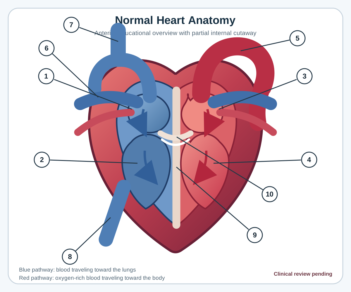
Numbered structure legend
| Number | Structure | Educational description |
|---|---|---|
| 1 | Right atrium | Receives blood returning from the body through the venae cavae. |
| 2 | Right ventricle | Receives blood from the right atrium. |
| 3 | Left atrium | Receives oxygen-rich blood returning from the lungs. |
| 4 | Left ventricle | Has a thick muscular wall and forms much of the heart apex. |
| 5 | Aorta | Begins above the left ventricle and arches upward. |
| 6 | Pulmonary arteries | The pulmonary trunk divides into right and left branches. |
| 7 | Superior vena cava | Enters the right atrium from above. |
| 8 | Inferior vena cava | Enters the lower portion of the right atrium. |
| 9 | Interventricular septum | Separates the right and left ventricles. |
| 10 | Heart valve region | Includes the tricuspid, pulmonary, mitral, and aortic valves. |
Viewer controls
- Switch between the normal 2D diagram and interactive 3D model.
- Zoom and reset the scalable normal diagram.
- Drag with a mouse or swipe to rotate the 3D model.
- Scroll or pinch to zoom the 3D model.
- Reset the 3D camera to its standard orientation.
- Enable optional automatic rotation.
- Open the anatomy viewer in fullscreen mode.
- Use the normal 2D diagram when 3D or WebGL is unavailable.
Three-dimensional asset provenance
The interactive model comes from NIH 3D entry 3DPX-022787, version 1.01. NIH 3D identifies the model as Public Domain. MaxArc Medical Library hosts a verified local GLB copy.
Current safety controls
- No diagnosis
- No prescribing
- No medication changes
- No clinical decision replacement
- No emergency-care replacement
Current 3D interaction boundary
The current NIH heart GLB is treated as a single-mesh model. Structure controls provide guided 3D camera orientations and synchronized educational descriptions.
True structure isolation, hiding, fading, removal, or mesh-level highlighting is not claimed. Those functions require a segmented, licensed, and clinically reviewed model.